Name: Human anatomy.
The authors: Prives M.G., Lysenkov N.K., Bushkovich V.I.
For more than 50 years, the textbook "Human Anatomy" has served higher medical education. Several generations of physicians began their journey into medicine by studying anatomy from this textbook.
In 1932, the first edition of the textbook "Human Anatomy", created by N.K. Lysenkov, was published. The fourth edition, which appeared in 1943, was prepared by V. I. Bushkovich. In 1958, the fifth edition of the textbook was published, in the preparation of which M. G. Prives took part. The fifth and all subsequent editions of the textbook (1968, 1969, 1974) were carried out by M. G. Prives. The eighth edition (1974) of the textbook "Human Anatomy" was awarded in 1981 with a diploma of the 1st degree of the Ministry of Health of the USSR as best textbook for higher medical schools.
The textbook has been repeatedly published in Spanish, and is currently being prepared for publication in English.
The present, ninth, edition is significantly revised and supplemented thanks to the great work of the Honored Scientist of the RSFSR Professor Mikhail Grigoryevich Prives, who from 1937 to 1977 headed the Department of Normal Anatomy of the 1st Leningrad Medical Institute. acad. I. P. Pavlova, and is currently its consultant professor.
The textbook is written taking into account the achievements of modern anatomical science. The material of the textbook is presented on the basis of the philosophy of dialectical materialism. Anatomy is presented not as a purely descriptive subject, but as an evolutionary, functional, effective and applied science - these are different aspects of one science - anatomy. New directions of anatomical science were also reflected - the influence of labor and sports on the structure of the human body. At the same time, individual variability is emphasized, due not only to genetic factors, but also to social ones.
The textbook examines the anatomy of a living person and emphasizes the differences in the structure of a living person from the structure and topography of organs on a corpse.
Human anatomy is presented not only in systems (systematic anatomy), that is, analytically, but also as a whole, which is in connection with its environment - synthetically. Therefore, at the end of the textbook, a synthesis of anatomical data is provided. Anatomical terms are aligned with the International Anatomical Nomenclature.
This edition of the textbook corresponds to the new curriculum on human anatomy, approved by the Ministry of Health of the USSR, and meets modern requirements for higher education textbooks.
Format: DjVu.
Pages: 672 pages
The year of publishing: 1985
Archive size: 12.4 MB.
Buy a book on human anatomy in Labyrinth.ru.
(born in 1904) - Soviet anatomist, doctor of medical sciences. Sciences (1937), Professor (1937), Honored. scientist of the RSFSR (1963). Member of the CPSU since 1939
He graduated in 1925 from medical school. Faculty of Voronezh University, worked there in the faculty surgical clinic, from 1930 to 1953 - in the State X-ray and Radiological Institute (now Ying t of Medical Radiology M3 USSR) in Leningrad; from 1937 to 1953 head. laboratory of normal p comparative anatomy in this in-those. At the same time (since 1937) professor, head. Department of human anatomy of the 1st Leningrad medical. in-ta, and since 1977 - professor-consultant of the same department. During the evacuation of the in-that to Krasnoyarsk (1942-1944) - one of the organizers and the first director of the Krasnoyarsk medical. in-ta.
M. G. Prives published approx. 200 scientific papers, including 5 monographs, has 6 copyright certificates. He studied the influence of human labor activity on changes in the structure of the musculoskeletal system and the vascular system; together with co-workers studied in animal experiments the adaptation of the vascular system to the conditions of space flight (gravitational overload, hypokinesia, physical inactivity, etc.). One of the first applied rentgenol. method for studying limf. system also received roentgenograms limf. vessels of the person in a wedge. conditions. He worked on the problem of collateral lymph circulation, regulating the influence of the nervous system on it, its condition under various extreme influences. M. G. Prives is the author of the formalin-free method of preserving corpses. They revised the textbook of anatomy by N. K. Lysenkov and V. I. Bushkovich, recommended for honey. universities of the USSR. This textbook has been translated into Spanish and has been published 4 times for Latin American countries. M. G. Prives was the first (since 1932) to read X-ray anatomy in the course of human anatomy.
M. G. Prives was elected chairman of the Leningrad branch of the All-Union Society of Anatomists, Histologists, Embryologists (honorary chairman since 1980), an honorary member of the All-Union and All-Russian Society of Anatomists, Histologists, Embryologists, as well as a number of foreign societies of anatomists (Mexico, Bulgaria, Czechoslovakia); was deputy head. editor of the journal "Archive of Anatomy, Histology and Embryology" (1950-1977).
He was awarded the Order of the Badge of Honor and medals.
Compositions: Blood supply of long tubular bones of the person, yew., L., 1938; Anatomy of intraorganic vessels, L., 1948 (author of a number of chapters and editors); Radiography of the lymphatic system, L., 1948; Methods of conservation of anatomical preparations, L., 1956; Human Anatomy, L., 1968, 1974 (jointly with others); Issues of aviation and space anatomy, c. 1, L., 1968 (author of a number of articles and editors); Further improvement of the method of preserving anatomical preparations, Arkh. aiat., gistol, and embryol., t. 58, no. 2, p. 96, 1970 (with others); Some results and perspectives of the cosmic anatomy of the vascular system, ibid., vol. 61, no. 11, p. 5, 1971; Biosocial problems of our time and anatomy, ibid., vol. 69, no. 10, p. 5, 1975; Influence various kinds sports on the growth of the skeleton in athletes of childhood, adolescence and youth, ibid., vol. 74, no. 6, p. 5, 1978 (jointly with Aleksina L.A.).
Bibliography: Mikhail Grigorievich Prives, Arch. anat., gistol, and embryol., t. 78, no. 3, p. 120, 1980.
N. V. Krylova.
This reissue was prepared by Doctor of Medical Sciences R.A. Prives-Bardina and Candidate of Medical Sciences O.M. Mikhailova. The terms in the textbook are given in accordance with the International Anatomical Nomenclature 2003.
Dear and dear students, future and already accomplished doctors and people who are simply interested in anatomy!
Before you is an amazing textbook, which in 2002 turned 70 years old. Over these long years, he has absorbed the wisdom of the authors who prepared him, and has been reprinted many times. Starting from the 5th edition, published in 1958, a person who is known as an outstanding scientist, an excellent teacher and a favorite of students, Professor Mikhail Grigorievich Prives, took part in its publication and editing.
This edition of the textbook is the 12th.
It is difficult to imagine that any textbook was reprinted 12 times in its Fatherland. Through the efforts and talent of Professor M.G. Prives, this textbook has turned from a guide to descriptive human anatomy into a reference book for many thousands of his students, absorbing all the new data on functional human anatomy, including the most modern scientific research conducted by students of Professor M.G. Weight gain according to the anatomy of a living person. This made human anatomy a science about a living person and for living people. This work also includes his well-known studies on X-ray anatomy and the anatomy of people of various professions, which he often liked to talk about: "terrestrial" and "unearthly", exposed to various factors of space flight. It is no coincidence that this textbook has been translated into various languages, including Spanish and English.
As a student of Mikhail Grigorievich, I am especially pleased to say a few kind words in the preface to the 12th edition, including because he dedicated the 10th edition to the 100th anniversary of the St. Petersburg Medical University. acad. I.P. Pavlov and the Department of Human Anatomy, where he worked for more than 60 years. Today, to our great regret, he is no longer with us, but the textbook, thought out and suffered by him, is reprinted once again, and this is the best memory of a person who will always live among us.
A person passes away, but the memory of him lives on in his deeds, is carefully kept in the minds and hearts of his students and all those who needed him and will always need him. That is why we bow our heads and kneel before the memory of talented person, A teacher with a capital letter, who is rightfully called and will be called the patriarch of Russian anatomy.
Head of the Department of Human Anatomy, St. Petersburg State Medical University. acad. I. P. Pavlova, Academician of the International Academy of Integrative Anthropology, Corresponding Member of the Petrovsky Academy of Sciences and Arts, Doctor of Medical Sciences, Professor A. Kosourov
- djvu format
- size 10.03 MB
- added October 27, 2010
9th ed. - M.: Medicine, 1985. - 672 p.
Textbook for medical students
For more than 50 years, the textbook "Human Anatomy" has served higher medical education. Several generations of doctors began their journey into medicine by studying anatomy using this textbook.
The textbook is written taking into account the achievements of modern anatomical science. The material of the textbook is presented on the basis of the philosophy of dialectical materialism. Anatomy is presented not as a purely descriptive subject, but as an evolutionary, functional, effective and applied science - these are different aspects of one science - anatomy. New directions of anatomical science were also reflected - the influence of labor and sports on the structure of the human body. At the same time, individual variability is emphasized, due not only to genetic factors, but also to social factors.
The textbook examines the anatomy of a living person and emphasizes the differences in the structure of a living person from the structure and topography of organs on a corpse.
Human anatomy is presented not only in systems (systematic anatomy), that is, analytically, but also as a whole, which is in connection with its environment - synthetically. Therefore, at the end of the textbook, a synthesis of anatomical data is provided. Anatomical terms are aligned with the International Anatomical Nomenclature
This edition of the textbook complies with the new human anatomy curriculum approved by the USSR Ministry of Health and meets modern requirements for higher education textbooks.
TABLE OF CONTENTS
Foreword
Introduction
Subject of anatomy (anatomy as a science)
Methods of anatomical research
A COMMON PART
Brief outline of the history of anatomy
Anatomy in Russia before the Great October Socialist Revolution
Anatomy in the USSR
General data on the structure of the human body
The body and its constituent elements
fabrics
Organs
Organ systems and devices
Body integrity
Organism and environment
The main stages of the individual development of the human body - onto
genesis
Extrauterine period of development of the organism
Human body shape, size, gender differences
The position of man in nature
The labor theory of F. Engels on the origin of man
Anatomical terminology
musculoskeletal system
Introduction
The passive part of the musculoskeletal system (the doctrine of bones and their joints -
osteoarthrology)
General osteology
Bone as an organ
Bone Development
Bone classification
The structure of bones in the x-ray image
Dependence of bone development on internal and external factors
General arthrology
Continuous connections - synarthroses
Discontinuous connections, joints, diarthrosis
Classification of joints and their general characteristics
Torso skeleton
vertebral column
Separate types of vertebrae
Connections between vertebrae
Connection of the vertebral column with the skull
Vertebral column as a whole
Rib cage
Sternum
Ribs
Rib connections
Chest as a whole
Head skeleton
Skull bones
Occipital bone
Sphenoid bone
Temporal bone
Parietal bone
frontal bone
Ethmoid bone
Bones of the face
upper jaw
palatine bone
Inferior turbinate
nasal bone
lacrimal bone
Coulter
Cheekbone
Lower jaw
Hyoid bone
Joints of the bones of the head
Skull as a whole
Age and sex features of the skull
Criticism of the racist "theory" in the doctrine of the skull (craniology)
limb skeleton
Phylogeny of limbs
Skeleton of the upper limb
Upper limb belt
Collarbone
shoulder blade
Joints of the bones of the girdle of the upper limb
Skeleton of the free upper limb
Brachial bone
shoulder joint
Forearm bones
Elbow bone
Radius
elbow joint
Connections of the bones of the forearm to each other
Hand bones
Wrist
metacarpus
Finger bones
Connections of the bones of the forearm with the hand and connections of the bones of the hand
Skeleton of the lower limb
Lower limb belt
Ilium
Pubic bone
Ischium
Joints of the pelvic bones
The pelvis as a whole
Skeleton of the free lower limb
Femur
hip joint
Patella
Lower leg bones
Tibia
Fibula
Knee-joint
Connections of the bones of the leg to each other
Foot bones
Tarsus
Metatarsus
Bones of the toes
Connections of the bones of the lower leg with the foot and between the bones of the foot
The active part of the musculoskeletal system (the study of muscles - myology)
General myology
Private myology
back muscles
Superficial back muscles
Deep back muscles
Autochthonous back muscles
Deep back muscles of ventral origin
Fascia of the back
Muscles of the ventral side of the body
chest muscles
Diaphragm
Breast fascia
Abdominal muscles
Neck muscles
Superficial muscles - derivatives of gill arches
Middle muscles, or muscles of the hyoid bone
deep muscles
Topography of the neck
Fascia of the neck
Muscles of the head
Chewing muscles
Facial muscles
Fascia of the head
Muscles of the upper limb
Muscles of the girdle of the upper limb
back group
front group
Shoulder muscles
Anterior shoulder muscles
Back muscles of the shoulder
Forearm muscles
front group
back group
Muscles of the hand
Fascia of upper limb and tendon sheath
Topography of the upper limb
Muscles of the lower limb
Muscles of the girdle of the lower limb
thigh muscles
Leg muscles
Foot muscles
Fascia of the lower limb and tendon sheath
Topography of the lower limb
The main specific features of the apparatus of human movement, which distinguish
him from animals
An overview of the muscles that produce the movements of the links of the body
THE DOCTRINE ABOUT THE INSIDE (SPLANCHNOLOGY). SPLANCHNOLOGIA
common data
Digestive system. Systema digestorium
Foregut derivatives
Oral cavity
Sky
Teeth
Language
Oral glands
Pharynx
Esophagus
Abdomen and pelvis
Stomach
Midgut derivatives
Small intestine
Hindgut derivatives
Colon
General patterns of intestinal structure
Large glands of the digestive system
Liver
Pancreas
Peritoneum
The main stages in the development of the digestive system and peritoneum and their anomalies
development
foregut
midgut
hindgut
Respiratory system. systema respiratorium
nasal cavity
Larynx
Trachea
Bronchi
Lungs
Pleural sacs and mediastinum
Development of the respiratory organs
Urogenital system. Systema urogenitale
urinary organs
Bud
Renal pelvis, cups and ureter
Bladder
female urethra
Sex organs. Organa genitalia
Male reproductive organs. Organa genitalia masculina
testicles
vas deferens
seminal vesicles
The spermatic cord and testicular membranes
Penis
male urethra
bulbourethral glands
Prostate
Female reproductive organs. Organa genitalia feminina
Ovary
Oviduct
Epididymis and periovary
Uterus
Vagina
female genital area
Development of the urinary organs
Crotch
TEACHER ABOUT THE ORGANIS OF INTERNAL SECRETION
Endocrine glands. Glandulae endocrinae
Branchiogenic group
Thyroid
parathyroid glands
Thymus
neurogenic group
Pituitary
Pineal body
Adrenal system group
Adrenal
Paraganglia
mesodermal glands
Endocrine parts of the gonads
Endodermal glands of the intestinal tube
Endocrine part of the pancreas
THE DOCTRINE ABOUT VESSELS (ANGIOLOGY). ANGIOLOGIA
Pathways that carry fluids
Circulatory system
Scheme of blood circulation
Development of the heart and blood vessels
Heart
chambers of the heart
The structure of the walls of the heart
Pericardium
Topography of the heart
Vessels of the small (pulmonary) circulation
Arteries of the small (pulmonary) circulation
Veins of the small (pulmonary) circulation
Vessels of the systemic circulation
Arteries of the systemic circulation
Aorta and its branches
Shoulder head trunk
common carotid artery
External carotid artery
internal carotid artery
subclavian artery
axillary artery
Brachial artery
radial artery
Ulnar artery
Branches of the descending aorta
Branches of the thoracic aorta
Branches of the abdominal aorta
Unpaired visceral branches
Paired visceral branches
Parietal branches of the abdominal aorta
internal iliac artery
External iliac artery
Arteries of the free lower limb
femoral artery
Popliteal artery
Anterior tibial artery
Posterior tibial artery
Arteries of the foot
Patterns of the distribution of arteries
Extraorgan arteries
Some patterns of branching of intraorgan arteries
Collateral circulation
Veins of the systemic circulation
Superior vena cava system
Brachiocephalic veins
Internal jugular vein
External jugular vein
Anterior jugular vein
subclavian vein
Veins of the upper limb
Veins - unpaired and semi-unpaired
Veins of the body walls
Vertebral plexus
Inferior vena cava system
Portal vein
Common iliac veins
Internal iliac vein
Porto-caval and cavo-caval anastomoses
External iliac vein
Veins of the lower limb
Patterns of distribution of veins
Features of the fetal circulation. ,
X-ray examination of blood vessels
lymphatic system
thoracic duct
Right lymphatic duct
Development of the lymphatic vessels
Lymphatic vessels and nodes of certain areas of the body
Patterns of distribution of lymphatic vessels and nodes
Collateral lymph flow
Organs of hematopoiesis and immune system
Spleen
STUDY OF THE NERVOUS SYSTEM (NEUROLOGY). SYSTEMA NERVOSUM
General data
Development of the nervous system
central nervous system
Spinal cord
Spinal cord retraction
Meninges of the spinal cord
Brain
General overview of the brain
brain embryogenesis
Separate parts of the brain
Rhomboid brain
Medulla
Hind brain
Bridge
Cerebellum
isthmus
IV ventricle
midbrain
forebrain
diencephalon
thalamic brain
Hypothalamus
III ventricle
telencephalon
Cloak
Olfactory brain
Lateral ventricles
Basal nuclei of the hemispheres
White matter of the hemispheres
Morphological bases of dynamic localization of functions in the cerebral cortex
large brain (centres of the cerebral cortex)
Falseness of the "theory" of racism in the doctrine of the brain
Shells of the brain
cerebrospinal fluid
Vessels of the brain
Peripheral part of the nervous system. zds
Animal or somatic nerves
spinal nerves
Posterior branches of the spinal nerves
Anterior branches of the spinal nerves
cervical plexus
Brachial plexus
Anterior branches of the thoracic nerves
Lumbosacral plexus
Lumbar plexus
sacral plexus
coccygeal plexus
cranial nerves
Nerves developed by fusion of spinal nerves
hypoglossal nerve
Gill arch nerves
Trigeminal nerve
facial nerve
Vestibulocochlear nerve
Glossopharyngeal nerve
Nervus vagus
accessory nerve
Nerves developing in connection with the head myotomes
oculomotor nerve
trochlear nerve
Abducens nerve
Nerves are derivatives of the brain
Olfactory nerves
optic nerve
Peripheral innervation of the soma
Patterns of distribution of nerves
Autonomic (autonomic) nervous system
Sympathetic part of the autonomic nervous system
Central part of the sympathetic part
Peripheral part of the sympathetic part
sympathetic trunk
Parasympathetic part of the autonomic nervous system
Parasympathetic centers
Peripheral division of the parasympathetic part
Brief overview of autonomic innervation of organs
The unity of the autonomic and animal parts of the nervous system
General overview of the main pathways of the nervous system
Diagram of the pathways of the nervous system
Afferent (ascending) pathways. §
Pathways from receptors of external stimuli
Pathways of the skin analyzer
Pathways from receptors of internal stimuli
Pathways of the motor analyzer
Interoceptive analyzer
The second afferent system of the brain - the reticular formation
Efferent (descending) pathways
Cortico-spinal (pyramidal) path, or pyramidal system
Descending pathways of the subcortical nuclei of the forebrain - extrapyramidal
system
Descending motor pathways of the cerebellum
Descending tracts of the cerebral cortex to the cerebellum
THE DOCTRINE ABOUT SENSE ORGANS (ESTHESIOLOGY). ORGAN SENSUUM
common data
Skin (organ of sense of touch, temperature and pain)
Milk glands
Predverio-y. peak organ
hearing organ
outer ear
Middle ear
inner ear
The organ of gravity and equilibrium (gravity analyzer, or stacokinetic analyzer
congestion)
Organ of vision
Eye
Eyeball
Shells of the eyeball
inner core of the eye
Accessory organs of the eye
organ of taste
Olfactory organ
THE PRINCIPLE OF INTEGRITY IN ANATOMY (SYNTHESIS OF ANATOMICAL
DATA)
see also

Crocker M. Human Anatomy
- pdf format
- size 12.76 MB
- added February 17, 2009
How does a person breathe? How do his eyes see? What happens to food when it enters the body? The answers to these and many other questions you can find in the book "Human Anatomy". Its author, Dr. Mark Crocker, presented detailed information in a lively and fascinating manner, accompanying the text with colorful illustrations and diagrams depicting human organs and systems. The book focuses on those that are...
Lecture number 1 - The human body as a whole. General patterns of growth and development
Presentation- ppt format
- size 1.12 MB
- added October 30, 2010
Department of Human and Animal Biology. Discipline "Anatomy and physiology of man". Introduction. Levels structural organization human body. The human body as a whole. General patterns of growth and development.
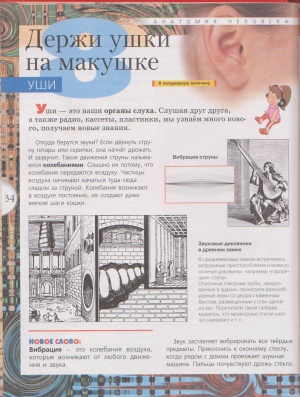
Mirer A.I. human anatomy
- jpg format
- size 25.05 MB
- added March 30, 2011
M.: Onyx Publishing House, 2008. - 88 p., ill. (Children's illustrated atlas) ISBN 978-5-488-01399-5 (1 design) ISBN 978-5-488-01596-8 (2 design) The book "Human Anatomy" is an additional guide to the courses "The World Around", "Biology "," Anatomy. It tells about how a person works, how our body works, and covers all issues and concepts of the general educational minimum established by the Ministry of Education and Science...

Presentations - Human Anatomy
Presentation- ppt format
- size 46.39 MB
- added March 05, 2011
In one archive, 4 voiced files of automated presentations on the topic "Human Anatomy" are combined: Parts of the body, 15 slides. Cells, skeleton, muscles, sensory organs, 18 slides. Brain, blood, circulatory system, lungs", 28 slides. Digestive system, glands, urinary and reproductive systems, development of the baby in the womb, 34 slides. Will be useful to educators, parents and teachers for developing individual and frontal activities ...
Name: Human anatomy.
For more than 50 years, the textbook "Human Anatomy" has served higher medical education. Several generations of physicians began their journey into medicine by studying anatomy from this textbook.
The textbook is written taking into account the achievements of modern anatomical science. The material of the textbook is presented on the basis of the philosophy of dialectical materialism. Anatomy is presented not as a purely descriptive subject, but as an evolutionary, functional, effective and applied science - these are different aspects of one science - anatomy. New directions of anatomical science were also reflected - the influence of labor and sports on the structure of the human body. At the same time, individual variability is emphasized, due not only to genetic factors, but also to social ones.
The textbook examines the anatomy of a living person and emphasizes the differences in the structure of a living person from the structure and topography of organs on a corpse.
Human anatomy is presented not only in systems (systematic anatomy), that is, analytically, but also as a whole, which is in connection with its environment - synthetically. Therefore, at the end of the textbook, a synthesis of anatomical data is provided. Anatomical terms are aligned with the International Anatomical Nomenclature.
This edition of the textbook complies with the new human anatomy curriculum approved by the USSR Ministry of Health and meets modern requirements for high school textbooks.
TABLE OF CONTENTS
Preface 3
Introduction 4
Subject of anatomy (anatomy as a science) 4
Methods of anatomical research 7
A COMMON PART
Brief outline of the history of anatomy 9
Anatomy in Russia before the Great October Socialist Revolution 14
Anatomy in the USSR 18
General data on the structure of the human body 20
Organism and its constituent elements 20
Fabrics 21
Organs 22
Organ systems and devices 23
Body integrity 25
Organism and environment 26
The main stages of the individual development of the human body - onto
genesis 27
Extrauterine period of development of the organism 32
Human body shape, size, gender differences 33
The position of man in nature 36
The labor theory of F. Engels on the origin of man 38
Anatomical terminology 40
musculoskeletal system
Introduction 43
The passive part of the musculoskeletal system (the doctrine of bones and their joints -
osteoarthrology) 44
General osteology 44
Bone as an organ 45
Bone development 47
Bone classification 51
The structure of bones in the x-ray image 52
Dependence of bone development on internal and external factors 55
General arthrology 58
Continuous connections - synarthroses 59
Discontinuous connections, joints, diarthrosis 61
Classification of joints and their general characteristics 63
Torso skeleton 56
Vertebral column 69
Separate types of vertebrae 70
Connections between vertebrae 76
Connection of the vertebral column with the skull 79
Vertebral column as a whole 80
Chest 82
Sternum 82
Ribs 82
Rib connections 83
Chest as a whole 84
Head skeleton 86
Skull bones 90
Occipital bone 90
Sphenoid bone 91
Temporal bone 92
Parietal bone 95
Frontal bone 96
Ethmoid bone 97
Facial bones 98
Upper jaw 98
Palatine bone 100
Inferior turbinate 101
Nasal bone 101
Lacrimal bone 101
Coulter 101
Cheekbone 102
Lower jaw 102
Hyoid bone 104
Joints of the bones of the head 105
Skull as a whole 107
Age and sex features of the skull 111
Criticism of the racist "theory" in the doctrine of the skull (craniology) 114
Skeleton limbs 115
Phylogeny of limbs 115
Upper limb skeleton 119
Upper limb belt 119
Clavicle 119
Blade 120
Connections of the bones of the girdle of the upper limb 121
Skeleton of the free upper limb 122
Humerus 122
Shoulder joint 123
Forearm bones 125
Ulna 125
Radius 125
Elbow joint 126
Connections of the bones of the forearm to each other 128
Hand bones 128
Wrist 128
Metacarpus 129
Finger bones 130
Connections of the bones of the forearm with the hand and connections of the bones of the hand 131
Lower limb skeleton 136
Lower limb belt 136
Ilium 136
Pubic bone 137
Ischium 137
Joints of the pelvic bones 138
Taz as a whole 139
Skeleton of the free lower limb 143
Femur 143
Hip joint 144
Patella 147
Leg bones 147
Tibia 147
Fibula 148
Knee joint 149
Connections of the bones of the lower leg to each other 152
Foot bones 153
Tarsus 153
Metatarsus 154
Bones of the toes 155
Connections of the bones of the lower leg with the foot and between the bones of the foot 156
The active part of the musculoskeletal system (the study of muscles - myology)
General myology 160
Private myology 169
Back muscles 169
Superficial back muscles 170
Deep back muscles 171
Autochthonous back muscles 171
Deep back muscles of ventral origin 174
Back fasciae 174
Muscles of the ventral side of the trunk 174
Chest muscles 175
Aperture 177
Breast fasciae 178
Abdominal muscles 178
Neck muscles 184
Superficial muscles - derivatives of gill arches 185
Middle muscles, or muscles of the hyoid bone 186
Deep muscles 188
Neck topography 189
Fascia of the neck 190
Muscles of the head 193
Chewing muscles 193
Facial muscles 193
Fascia of the head 196
Muscles of the upper limb 197
Muscles of the girdle of the upper limb 197
Rear group 197
Front group 200
Shoulder muscles 200
Anterior shoulder muscles 200
Back muscles of the shoulder 201
Muscles of the forearm 201
Front group 202
Rear group 203
Muscles of the hand 207
Fascia of upper limb and tendon sheath 210
Topography of the upper limb 212
Muscles of the lower limb 214
Muscles of the girdle of the lower limb 214
Muscles of the thigh 217
Calf muscles 220
Muscles of the foot 224
Fascia of the lower limb and tendon sheath 227
Topography of the lower limb 230
The main specific features of the apparatus of human movement, which distinguish
its from animals 232
An overview of the muscles that produce the movements of the links of the body 233
THE DOCTRINE OF THE INSIDE (SPLANCHNOLOGY) SPLANCHNOLOGIA
General data 235
Digestive System Systema digestorium 237
Foregut derivatives 237
Oral cavity 237
Sky 238
Teeth 240
Language 250
Oral glands 253
Throat 255
Esophagus 257
Abdomen and pelvis 260
Stomach 262
Midgut derivatives 269
Small intestine 269
Hindgut derivatives 275
Large intestine 275
General laws of the structure of the intestine 282
Large glands of the digestive system 283
Liver 283
Pancreas 288
Peritoneum 289
The main stages of development of the digestive system and peritoneum and anomalies of their development 295
Foregut 297
Midgut 298
Hind gut 298
Respiratory system Systema respiratorium 300
Nasal cavity 301
Larynx 303
Trachea 308
Bronchi 309
Lungs 309
Pleural sacs and mediastinum 316
Development of the respiratory organs 319
Genitourinary system Systema urogenitale 321
Urinary organs 322
Kidney 322
Renal pelvis, cups and ureter 326
Bladder 330
Female urethra 332
Genital organs Organa genitalia 333
Male reproductive organs Organa genitalia masculina 333
Testicles 333
Deferent duct 335
Seminal vesicles 335
The spermatic cord and testicular membranes 336
Penis 339
Male urethra 340
Bulbourethral glands 343
Prostate gland 343
Female reproductive organs Organa genitalia feminina 345
Ovary 345
Fallopian tube 347
Epididymis and periovary 347
Uterus 347
Vagina 352
Female genital area 353
Development of the urinary organs 355
Crotch 357
TEACHER ABOUT THE ORGANIS OF INTERNAL SECRETION
Endocrine glands Glandulae endocrinae 363
Branchiogenic group 365
Thyroid gland 365
Parathyroid glands 367
Thymus gland 367
Neurogenic group 368
Pituitary 368
Pineal body 370
Adrenal system group 370
Adrenal 370
Paraganglia 373
Mesodermal glands 373
Endocrine parts of the gonads 373
Endodermal glands of the intestinal tube 374
Endocrine part of the pancreas 374
THE DOCTRINE ABOUT VESSELS (ANGIOLOGY) ANGIOLOGIA
Paths carrying fluids 375
Circulatory system 376
Circulation scheme 378
Development of the heart and blood vessels 381
Heart 385
Chambers of the heart 387
The structure of the walls of the heart 390
Pericardium 397
Topography of the heart 398
Vessels of the small (pulmonary) circulation 402
Arteries of the small (pulmonary) circulation 402
Veins of the small (pulmonary) circulation 403
Vessels of the systemic circulation 403
Arteries of the systemic circulation 403
Aorta and branches of its arch 403
Shoulder stem 404
Common carotid artery 404
External carotid artery 404
Internal carotid artery 407
Subclavian artery 409
Axillary artery 412
Brachial artery 414
Radial artery 415
Ulnar artery 415
Branches of the descending aorta 418
Branches of the thoracic aorta 418
Branches of the abdominal aorta 418
Unpaired visceral branches 418
Paired visceral branches 421
Parietal branches of the abdominal aorta 422
Internal iliac artery 422
External iliac artery 424
Arteries of the free lower limb 425
Femoral artery 425
Popliteal artery 426
Anterior tibial artery 427
Posterior tibial artery 428
Foot arteries 428
Patterns of the distribution of arteries 430
Extraorgan arteries 430
Some patterns of branching of intraorgan arteries 432
Collateral circulation 434
Veins of the systemic circulation 436
Superior vena cava system 436
Brachiocephalic veins 436
Internal jugular vein 436
External jugular vein 438
Anterior jugular vein 439
Subclavian vein 439
Veins of the upper limb 439
Veins - unpaired and semi-unpaired 441
Veins of the walls of the body 442
Vertebral plexuses 442
Inferior vena cava system 442
Portal vein 443
Common iliac veins 445
Internal iliac vein 445
Porto-caval and cavo-caval anastomoses 446
External iliac vein 447
Veins of the lower limb 447
Patterns of distribution of veins 448
Features of the fetal circulation, 449
X-ray examination of blood vessels 451
Lymphatic system 454
Thoracic duct 458
Right lymphatic duct 458
Development of lymphatic vessels 459
Lymphatic vessels and nodes of certain areas of the body 460
Patterns of distribution of lymphatic vessels and nodes 467
Collateral lymph flow 468
Organs of hematopoiesis and immune system 470
Spleen 470
STUDY OF THE NERVOUS SYSTEM (NEUROLOGY) SYSTEMA NERVOSUM
General information 473
Development of the nervous system 477
Central nervous system 482
Spinal cord 482
Spinal cord resection 483
Meninges of the spinal cord 489
Brain 491
General overview of the brain 491
Embryogenesis of the brain 493
Separate parts of the brain 496
Rhomboid brain 497
medulla oblongata 497
Hind brain 500
Bridge 500
Cerebellum 502
Isthmus 504
IV ventricle 504
Midbrain 508
Forebrain 511
Diencephalon 511
Thalamic brain 511
Hypothalamus 513
III ventricle 514
telencephalon 515
Cloak 516
Olfactory brain 521
Lateral ventricles 522
Basal nuclei of the hemispheres 523
White matter of the hemispheres 526
Morphological bases of dynamic localization of functions in the cerebral cortex
large brain (centers of the cerebral cortex) 528
The Falsity of the "Theory" of Racism in the Doctrine of the Brain 537
Meninges 538
Cerebrospinal fluid 542
Vessels of the brain 543
Peripheral part of the nervous system
Animal or somatic nerves 545
Spinal nerves 545
Posterior branches of spinal nerves 545
Anterior branches of spinal nerves 546
Cervical plexus 547
Brachial plexus 548
Anterior branches of the thoracic nerves 552
Lumbosacral plexus 553
Lumbar plexus 553
sacral plexus 554
Coccygeal plexus 558
Cranial nerves 558
Nerves developed by fusion of spinal nerves 560
Hypoglossal nerve 560
Gill arch nerves 562
Trigeminal nerve 563
Facial nerve 569
Vestibulocochlear nerve 572
Glossopharyngeal nerve 573
Vagus nerve 574
Accessory nerve 578
Nerves developing in connection with head myotomes 578
Oculomotor nerve 578
trochlear nerve 579
Abducens nerve 579
Nerves are derivatives of the brain 579
Olfactory nerves 579
Optic nerve 579
Peripheral innervation of the soma 582
Patterns of distribution of nerves 585
Autonomic (autonomic) nervous system 586
Sympathetic part of the autonomic nervous system 593
Central division of the sympathetic part 593
Peripheral division of the sympathetic part 593
Sympathetic trunk 594
Parasympathetic part of the autonomic nervous system 597
Centers of the parasympathetic part 597
Peripheral division of the parasympathetic part 598
Brief overview of the autonomic innervation of organs 599
The unity of the autonomic and animal parts of the nervous system 503
General overview of the main pathways of the nervous system 605
Diagram of the pathways of the nervous system 607
Afferent (ascending) pathways §07
Pathways from receptors of external stimuli 507
Pathways of the skin analyzer 507
Pathways from receptors of internal stimuli 510
Pathways of the 510 Motor Analyzer
Interoceptive Analyzer 512
The second afferent system of the brain - the reticular formation 514
Efferent (descending) pathways 615
Cortico-spinal (pyramidal) path, or pyramidal system 616
Descending pathways of the subcortical nuclei of the forebrain - extrapyramidal
system 616
Descending motor pathways of the cerebellum 618
Descending pathways of the cerebral cortex to the cerebellum 618
THE DOCTRINE ABOUT THE SENSE ORGANS (ESTHESIOLOGY) ORGANA SENSUUM
General data 620
Skin (organ of touch, temperature and pain) 622
Mammary glands 624
Vestibule-u-peak organ 625
Organ of hearing 627
Outer ear 627
Middle ear 629
Inner ear 632
Organ of gravity and balance (gravity analyzer, or stacokinetic analyzer) 638
Organ of vision 640
Eye 641
Eyeball 641
Shells of the eyeball 641
Inner nucleus of the eye 647
Auxiliary organs of the eye 648
Organ of taste 653
Olfactory organ 654
THE PRINCIPLE OF INTEGRITY IN ANATOMY (SYNTHESIS OF ANATOMICAL
DATA)